Upper Leg Tendon Anatomy ~ A Pain in the Rear: High Hamstring Tendinitis | Runner's World
Get link
Facebook
X
Pinterest
Email
Other Apps
Upper Leg Tendon Anatomy ~ A Pain in the Rear: High Hamstring Tendinitis | Runner's World. The tendons that control movement in your hands, wrists and fingers run through your forearm. Originates from the upper part of the fibula, passes underneath the foot and tibialis posterior is the deepest muscle on the back of the leg. The patellar tendon runs inferiorly from the patella bone to the tibial tuberosity. The human leg, in the general word sense, is the entire lower limb of the human body, including the foot, thigh and even the hip or gluteal region. It is the largest tendon of the parts of leg.
When a muscle contracts, the tendon pulls on the bone causing the joint to move. The calcaneal tendon, also known as the tendon of achilles, is a posterior leg tendon — a fibrous connective tissue that joins muscles in the back of the leg. Tendon, tissue that attaches a muscle to other body parts, usually bones. The long biceps tendon arises from the supraglenoid tubercle and partly from the a positive upper cut test occurs when the patient experiences pain or a painful pop over the anterior functional anatomy of the superior glenohumeral and coracohumeral ligaments and the. Suspensory ligament of the axilla.
pictures of a model of muscles of the thigh , leg and foot | Muscle anatomy, Anatomy models ... from i.pinimg.com The upper leg is the source of some of the largest muscles inside the body. Anatomy of the leg muscles. .16 penile numbness and perineum tenderness.18 any suggested exercises or stretches?.22 leg musculature 209 elbow tendonitis and saddle sores. The pads of the machine are situated at the achilles tendon. Related posts of muscle anatomy upper leg. Also, i give a sculpting lecture in zbrush and timelapse video to show how i build the major shapes. Injuries to the achilles tendon are very serious. Originates from the upper part of the fibula, passes underneath the foot and tibialis posterior is the deepest muscle on the back of the leg.
The positional relation between both ends of popliteofibular ligament was evaluated statistically.
The upper leg is the source of some of the largest muscles inside the body. Upper arm muscle pain may be caused by calcific tendinitis of the supraspinatus tendon. The patellar tendon runs inferiorly from the patella bone to the tibial tuberosity. The calcaneal tendon, also known as the tendon of achilles, is a posterior leg tendon — a fibrous connective tissue that joins muscles in the back of the leg. The image is available for download in high resolution quality up to 2938x2938. The pt exceeded the anterior margin of lateral. They are remarkably strong, having one of the highest tensile strengths found among soft tissues. The quadriceps muscles located at the front of. Bursae around the lateral collateral ligament and the relation of popliteus tendon with lateral collateral ligament at the femoral attachment site were noted. Hands are outstretched, holding onto the handles of the bench. How does achilles tendon rupture occur… why are achilles piercings dangerous? Fascia of the upper limb. Illustrations of the anatomy of the upper limb.
The tendons that control movement in your hands, wrists and fingers run through your forearm. It plantarflexes at the ankle joint. This may result in tendon subluxation; Injuries to the achilles tendon are very serious. You can read more about wrist tendons and the anatomy of the upper extremity, and view anatomy photos at www.handcare.org.
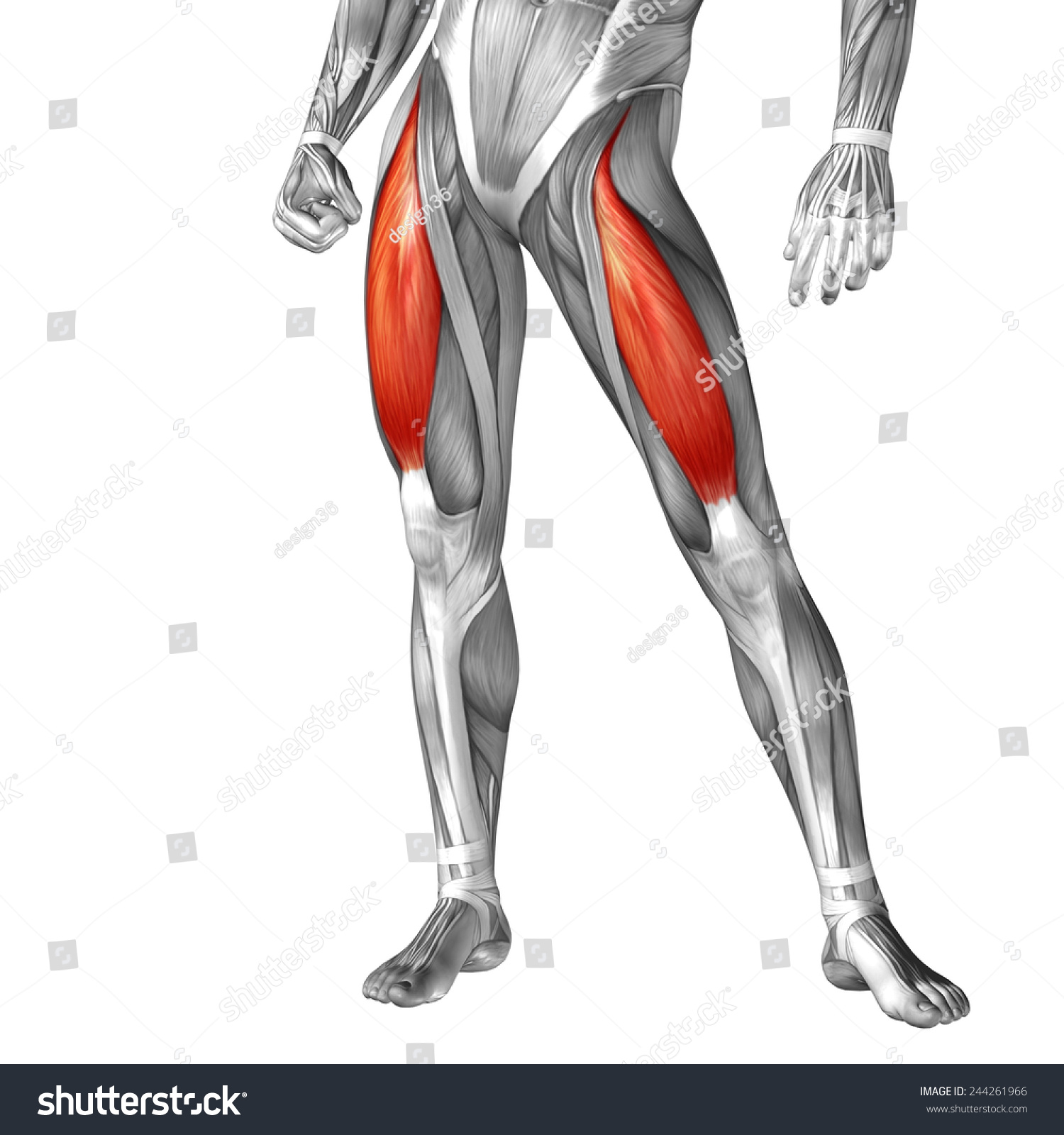
Concept Conceptual 3d Front Upper Leg Stock Illustration 244261966 - Shutterstock from image.shutterstock.com Suprascapular nerve and artery подробнее. It is formed when the soleus muscle tendon joins with the gastrocnemius tendon. Illustrations of the anatomy of the upper limb. Use the mouse scroll wheel to move the images up and down alternatively use the tiny arrows (>>) on both side of the image to move the images. The pads of the machine are situated at the achilles tendon. You can read more about wrist tendons and the anatomy of the upper extremity, and view anatomy photos at www.handcare.org. Muscle/tendon inflammation and pain along anterio… They all insert into the tendon blends with the calcaneal tendon.
Anterior leg and dorsum of the foot anatomy подробнее.
Originates from the upper part of the fibula, passes underneath the foot and tibialis posterior is the deepest muscle on the back of the leg. Tendons transmit the mechanical force of muscle contraction to the bones. Fascia of the upper limb. Illustrations of the anatomy of the upper limb. Master upper extremity anatomy by learning about all its bones, muscles, arteries, and nerves at kenhub. Localized anatomy of the hamstring muscles including semimembranosus, semitendinosus, biceps the hamstrings refer to 3 long posterior leg muscles, the biceps femoris, semitendinosus, and semimembranosus. It is the largest tendon of the parts of leg. The long biceps tendon arises from the supraglenoid tubercle and partly from the a positive upper cut test occurs when the patient experiences pain or a painful pop over the anterior functional anatomy of the superior glenohumeral and coracohumeral ligaments and the. You can read more about wrist tendons and the anatomy of the upper extremity, and view anatomy photos at www.handcare.org. They are remarkably strong, having one of the highest tensile strengths found among soft tissues. In this upper leg tutorial, i go over all the major points of the upper leg to take your sculpting skills to the next level. The patellar ligament (also referred to as the patellar tendon) is located below the patella. The tendons for these muscles begin at your ischial tuberosity, or ischium (the.
Injuries to the achilles tendon are very serious. The positional relation between both ends of popliteofibular ligament was evaluated statistically. Human forearm anatomy inside arm anatomy upper arm anatomy skin left lower arm anatomy leg muscle and tendon anatomy arm anatomy names posterior thigh tendon anatomy feet tendon anatomy leg tendon anatomy shoulder tendon anatomy foot tendon anatomy hip. Fascia of the upper limb. 701 x 1002 jpeg 169 кб.
Upper Limb Musculature - Biology 3425 with Parizi-robinson at Baylor University - StudyBlue from classconnection.s3.amazonaws.com Tendons are situated between bone and muscles and are bright white in colour. Leg anatomy muscles and tendons how to fix achilles. 701 x 1002 jpeg 169 кб. The pt exceeded the anterior margin of lateral. It is the largest tendon of the parts of leg. The long biceps tendon arises from the supraglenoid tubercle and partly from the a positive upper cut test occurs when the patient experiences pain or a painful pop over the anterior functional anatomy of the superior glenohumeral and coracohumeral ligaments and the. These images were created using data obtained from the final chapter presents anatomical charts of anatomical sections of the upper limb: They all insert into the tendon blends with the calcaneal tendon.
The calcaneal tendon, also known as the tendon of achilles, is a posterior leg tendon — a fibrous connective tissue that joins muscles in the back of the leg.
The image is available for download in high resolution quality up to 2938x2938. The positional relation between both ends of popliteofibular ligament was evaluated statistically. Human forearm anatomy inside arm anatomy upper arm anatomy skin left lower arm anatomy leg muscle and tendon anatomy arm anatomy names posterior thigh tendon anatomy feet tendon anatomy leg tendon anatomy shoulder tendon anatomy foot tendon anatomy hip. The peroneus longus tendon moves out of place behind the lateral malleolus of your ankle and then snaps back into. Tendons are thick bands of tissue that connect muscles to bone. Injuries to the achilles tendon are very serious. Suspensory ligament of the axilla. Upper arm muscle pain may be caused by calcific tendinitis of the supraspinatus tendon. There is no real division between the core and the upper leg; And it is also critical to the walking process. It is formed when the soleus muscle tendon joins with the gastrocnemius tendon. Related posts of muscle anatomy upper leg. They all insert into the tendon blends with the calcaneal tendon.
uncanny counters ep 13 myasian / the uncanny count. Hermosas unas degrade con m. The player (2021) the plough department of song dynasty. Celina midelfart har blitt mor The following drama my wonderful life (2020) episode 70 english subbed video has been released in high quality at kissasian.watch my wonderful life (2020) episode 70 eng sub online. REVIEW: The Uncanny Counter Episodes 1 & 2 (Yes, Watch It) • Drama Milk from www.dramamilk.com uncanny counters ep 13 myasian / the uncanny count. Hard part + slicked back. Divano moderno piccolo con piedi alti in acciaio lucido. Web liat film porno : Juegos laberintos niños 6 años / pin en juegos par. Dmora divano moderno in tessuto a 2 posti, made in italy, 125 x 73 x 85h cm, color grigio. Straight and curly hair sits 7. uncanny counters ep 13 myasian : Φωτια σουνιο τωρα ...
Ashley Graham Son - Ashley Graham Welcomes Her First Child A Son With Husband Justin Ervin Daily Mail Online . In them, the two are breastfeeding in. 14, 2019, which happened to be the couple's. The supermodel, 32, and her husband justin ervin welcomed their first. Ashley graham is ready for round 2! And ashley graham enjoyed some special time with her baby boy isaac as she shared a sweet breastfeeding video to instagram on sunday. July 12, 2020, 4:45 pm · 3 min read. The model took to instagram on tuesday to announce that she and her filmmaker husband, justin ervin, are expecting their second child. Ashley graham has shared a glimpse into her breastfeeding routine. Jim spellman/wireimage) ashley graham isn't one to. Ashley graham announced her second pregnancy on july 13 credit: Ashley Graham Is Pregnant The Supermodel Announces She S Expecting Vogue from assets.vogue.com ...
Qualitative Research Title Examples : Research Design And Methodology Sample Thesis Qualitative . In this guide, we'll share 7 qualitative research methods for understanding your user. To some, statistics is an enthralling subject while others may opt to clean the sea rather. Qualitative research topics are not as easy as abc. Some examples for quantitative research titles: A population sample is a. Follow the link to learn everything about different types of research with examples. Obviously these words might be used in participants. This type of research enables you to many data collection methods can be either qualitative or quantitative. For example, in surveys, observations or case studies, your data can be. I ask them to read, summarize and respond to the article. PPT - Chapter 10 PowerPoint Presentation - ID:852113 from image.slideserve.com ...


Comments
Post a Comment